Retinal Detachment
Retinal detachment occurs when the retina separates from its underlying supporting tissue and is a serious eye condition that requires urgent evaluation.

Prof. Dr. Fevzi Şentürk
Ophthalmology · Istanbul
▶ Retina Hastalıkları ve Tedavi Yöntemleri (CNN Türk Haber)
{ AI · preliminary guidance }
onlineLet's talk about your retina
AI responses do not replace a medical diagnosis.
Konuyla ilgili kısa videolar
Tüm videolar →Kliniğin YouTube kanalından bu konuyla ilgili kısa açıklamalar. Toplam 3 video.

Vitrektomi Nedir ve Nasıl Tedavi Edilir?

SSR Hastalığı ve Fotodinamik Tedavi Nedir?

Tavuk Karası (Retinitis Pigmentosa) Hastalığı Nedir?
Retinal detachment is an emergency condition in which the retina separates from its normal position, and it can lead to permanent vision loss if left untreated.
What Is Retinal Detachment?
Retinal detachment is the separation of the retina — the light-sensitive tissue at the back of the eye — from its underlying supporting tissue, the choroid. Once separated, the retina no longer receives adequate nourishment and oxygen, which can rapidly impair its function. Retinal detachment is a serious eye condition that requires urgent evaluation and, in most cases, urgent treatment.
What Are the Types of Retinal Detachment?
Rhegmatogenous Retinal Detachment
This is the most common type. It develops when fluid passes through a tear or hole in the retina and accumulates beneath it. It is often associated with age-related vitreous changes, high myopia, or trauma.
Tractional Retinal Detachment
This occurs when scar tissue on the retinal surface pulls the retina away from its position. It can be seen in conditions such as advanced diabetic retinopathy.
Exudative Retinal Detachment
This type is characterised by fluid accumulation beneath the retina without a tear or hole. It may be associated with certain inflammatory or vascular conditions.
What Are the Symptoms of Retinal Detachment?
- Sudden flashes of light (photopsia)
- A marked and sudden increase in floaters
- A curtain, shadow, or dark-area sensation over part of the visual field
- Sudden and progressive blurring of vision
If any of these symptoms occur, prompt consultation with an ophthalmologist is recommended.
Who Is More Likely to Develop Retinal Detachment?
The following factors can increase the risk of retinal detachment:
- High myopia (strong glasses prescription)
- Previous cataract surgery
- History of eye trauma
- Family history of retinal detachment
- Advanced diabetic retinopathy
- Previously identified weak areas or tears in the retina
How Is Retinal Detachment Diagnosed?
Diagnosis is made through a dilated eye examination. When needed, additional imaging methods such as ultrasonography may be used, particularly in cases where vitreous haemorrhage or other factors make direct visualisation difficult.
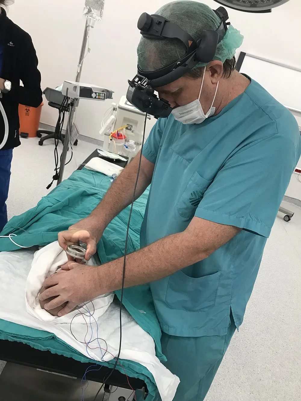
How Is Retinal Detachment Treated?
The treatment method is determined based on the type, extent, and duration of the detachment, and whether the macula is involved.
Pneumatic Retinopexy
A gas bubble is injected into the eye to help reposition the retina; the tear is usually supported with laser or cryotherapy.
Scleral Buckling
A silicone band is placed on the outside of the eye wall to help bring the retina back into contact with its underlying supporting tissue.
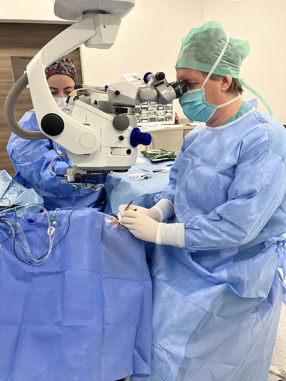
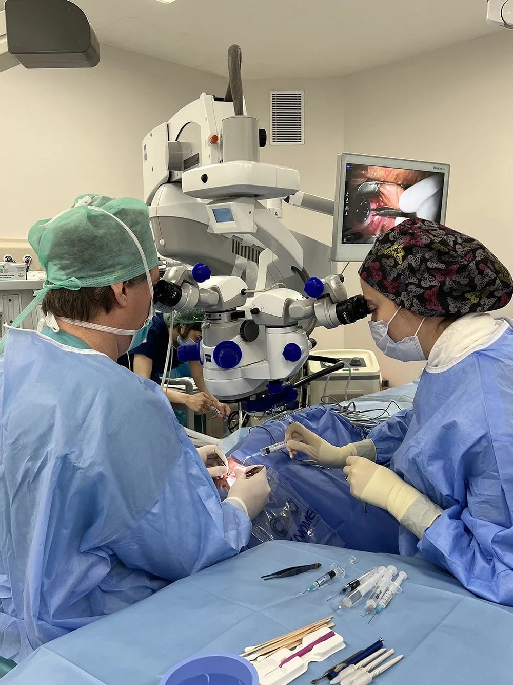
Vitrectomy Surgery
The vitreous gel inside the eye is removed to allow the retina to be repositioned and flattened. A temporary gas bubble or silicone oil tamponade may be left in the eye at the end of the procedure.
The choice of method is determined by the doctor based on the patient’s clinical condition.
What Is the Recovery Process Like After Retinal Detachment Treatment?
- A specific head positioning may need to be maintained for a period after treatment, especially when a gas tamponade is used
- Visual clarity may improve gradually; full recovery can take weeks to months
- Air travel should not be undertaken without a doctor’s approval while a gas bubble remains in the eye
- Regular follow-up examinations are necessary to monitor the healing process
What Happens if Retinal Detachment Is Left Untreated?
Untreated retinal detachment can lead to progressive and often permanent vision loss in the affected area of the retina. Early recognition of symptoms and urgent evaluation directly affect the chance of preserving vision.
Frequently Asked Questions
What is retinal detachment?
Retinal detachment is the separation of the retina — the light-sensitive tissue at the back of the eye — from its underlying supporting tissue. This disrupts normal retinal function and requires urgent evaluation.
What are the symptoms of retinal detachment?
The most common symptoms are sudden flashes of light, a marked increase in floaters, a curtain- or shadow-like sensation over part of the visual field, and sudden blurring of vision.
Is retinal detachment an emergency?
Yes. If retinal detachment is suspected, an ophthalmologist should be consulted without delay. Early intervention increases the chance of preserving vision.
Who is more likely to develop retinal detachment?
People with high myopia, those who have previously had cataract surgery, those with a history of eye trauma, those with a family history of retinal detachment, and those with advanced diabetic retinopathy are at higher risk.
What are the types of retinal detachment?
The most common type is rhegmatogenous retinal detachment, caused by a tear or hole in the retina. Tractional and exudative retinal detachment can also occur.
How is retinal detachment treated?
The treatment approach depends on the type, extent, and duration of the detachment. Pneumatic retinopexy, scleral buckling, and vitrectomy surgery are the main treatment methods.
Does vision return after retinal detachment surgery?
The degree of visual recovery depends on the duration of the detachment, whether the macula was involved, and how promptly treatment was started. Early treatment is generally associated with better outcomes.
What happens if retinal detachment is left untreated?
Untreated retinal detachment can lead to progressive and permanent vision loss. This makes urgent evaluation essential once symptoms are noticed.
Is it possible to prevent retinal detachment?
Regular dilated eye examinations for people with high myopia and other at-risk individuals can help detect retinal tears early, allowing preventive treatment, such as laser, before a detachment develops.
Treatments often evaluated together
-
Vitrectomy
Vitrectomy is a surgical procedure in which the eye's vitreous gel is removed, used to treat a range of retinal conditions such as retinal detachment, complications of diabetic retinopathy and macular hole.
-
Intravitreal Injection
Intravitreal injection is a procedure used to treat retinal diseases such as macular degeneration, diabetic macular oedema and retinal vein occlusion, in which medication is delivered directly into the eye.
-
Cataract Surgery
Cataract surgery is the procedure in which the eye's natural lens, clouded by a cataract, is removed and replaced with an artificial intraocular lens (IOL) to correct blurred vision.


Book an Appointment
Your information reaches Prof. Dr. Fevzi Şentürk's clinic. A response is made within 24 hours.